

 in Da Nang with Dr. Dang Phuoc Dat, MD, Specialist Level I (+84 777 743 777)—the EAU and AUA gold standard with <1% recurrence and maximum testicular artery and lymphatic preservation.)
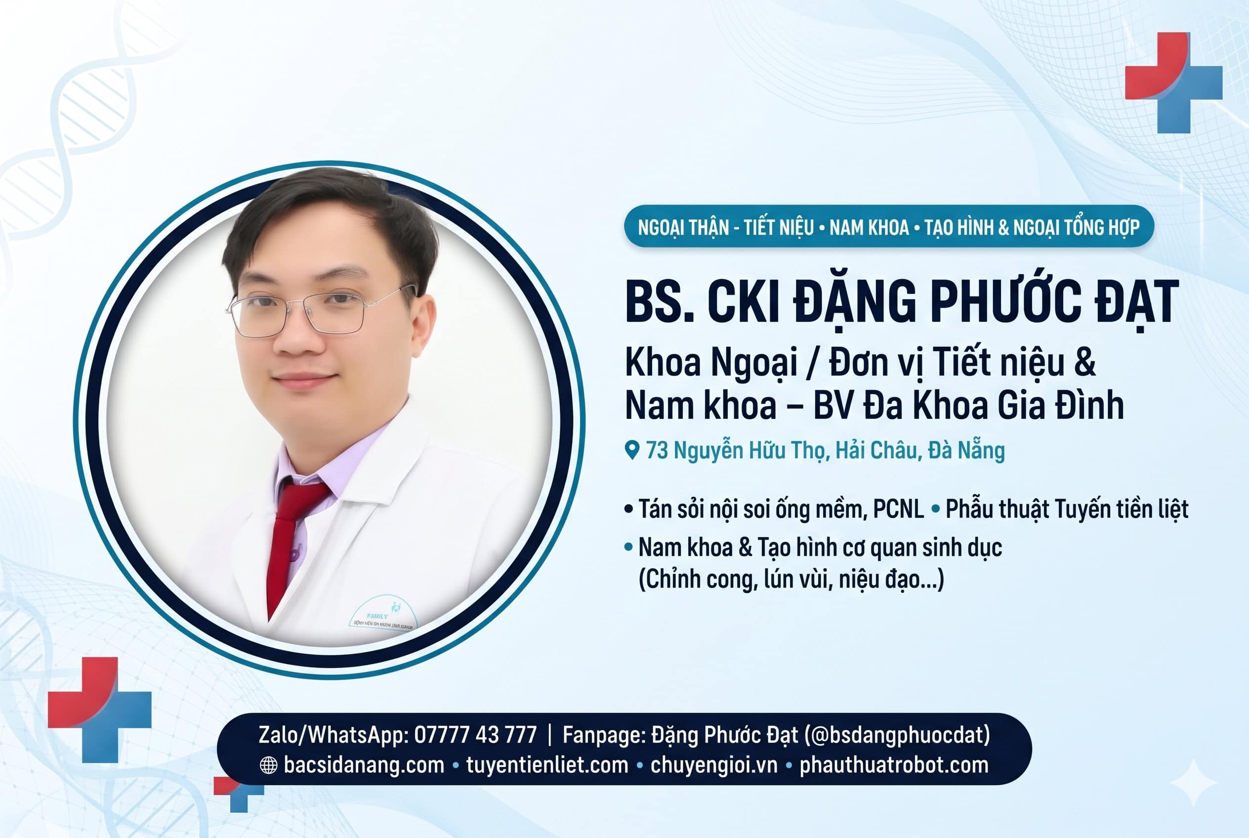
Author & Medical Reviewer: Dr. Dang Phuoc Dat, MD, Specialist Level I – Consultant Urologist, Andrologist & Reconstructive Surgeon (Family Hospital Da Nang & 162 Te Hanh Private Clinic)
Clinical Executive Summary: Clinical varicocele is the most common correctable cause of male infertility worldwide, present in 35%–40% of men with primary infertility and up to 80% of men with secondary infertility. Both the European Association of Urology (EAU 2026) and American Urological Association (AUA/ASRM) guidelines designate Subinguinal Microsurgical Varicocelectomy (Marmar Technique) as the undisputed surgical gold standard—improving sperm concentration and motility in 65%–75% of patients while reducing postoperative hydrocele and recurrence rates below 1%.
1. How Varicocele Impairs Male Fertility, Testosterone, and Scrotal Comfort
A varicocele is an abnormal dilation and tortuosity of the internal spermatic veins within the pampiniform plexus draining the testicle—occurring predominantly on the left side (85%–90%) due to the perpendicular insertion of the left gonadal vein into the high-pressure left renal vein [1, 2]. When incompetent venous valves allow retrograde blood reflux down into the scrotum, three pathophysiological cascades damage spermatogenesis and Leydig cell function:
- Scrotal Hyperthermia (Loss of Countercurrent Heat Exchange): Normal spermatogenesis requires intrascrotal temperatures 1.5°C to 2.5°C cooler than core body temperature. Pooling of warm venous blood around the testis elevates testicular temperature, arresting spermatid maturation.
- Oxidative Stress & Elevated Sperm DNA Fragmentation Index (DFI): Venous stasis increases reactive oxygen species (ROS), causing single- and double-stranded breaks in sperm chromatin—linked to recurrent miscarriage and IVF/ICSI failure [1].
- Leydig Cell Dysfunction & Subclinical Hypogonadism: Chronic intratesticular venous hypertension impairs testosterone biosynthesis, while stretching of perivascular nerve fibers causes a characteristic dull, dragging scrotal ache that worsens after prolonged standing or gym workouts.
CLINICAL VISUAL DIAGRAM: DUBIN & AMELAR VARICOCELE GRADING & COLOUR DOPPLER CRITERIA
| Clinical Grade | Physical Examination (Standing Position) | Scrotal Colour Doppler Ultrasound Findings |
|---|---|---|
| Grade I (Small Clinical) | Palpable only during a forceful Valsalva maneuver while standing. | Maximal venous diameter 2.5 – 3.0 mm with retrograde reflux > 2 seconds on Valsalva. |
| Grade II (Moderate Clinical) | Easily palpable at rest while standing without Valsalva, but not visible through scrotal skin. | Dilated pampiniform veins 3.1 – 4.0 mm with continuous venous reflux during Valsalva. |
| Grade III (Large “Bag of Worms”) | Clearly visible through the scrotal skin at rest (“bag of worms” deformity), often with ipsilateral testicular hypotrophy. | Tortuous venous plexus > 4.0 mm at rest with spontaneous or marked reflux and reduced testicular volume (>20% asymmetry). |
2. Why Subinguinal Microsurgical Varicocelectomy (Marmar Technique) Is the Gold Standard
Introduced by Dr. Joel Marmar and refined with high-power optical magnification, Subinguinal Microsurgical Varicocelectomy accesses the spermatic cord through a miniature 2.0 cm to 2.5 cm incision just below the external inguinal ring—without cutting any abdominal fascia or muscle fibers [2, 3].
According to systematic reviews published in World Journal of Men's Health (NCBI PMC – Clinical Outcomes of Varicocele Repair) and the WHO & EAU Male Infertility Guidelines (NCBI Bookshelf), performing the dissection under 10x to 20x microsurgical magnification allows Dr. Dang Phuoc Dat, MD, Specialist Level I to achieve three anatomical objectives impossible with macroscopic or laparoscopic surgery:
- Complete Ligation of Internal and External Spermatic Veins: Eliminates all venous reflux pathways (reducing recurrence to < 1% – 1.5%, compared to 10%–15% with non-microsurgical techniques).
- Pinpoint Identification & Preservation of the Testicular Artery (1.0 mm diameter): Prevents accidental arterial ligation, protecting testicular volume and Leydig cell testosterone output.
- Preservation of Transparent Lymphatic Channels: Prevents postoperative tunica vaginalis fluid accumulation, reducing the risk of secondary postoperative hydrocele to nearly 0% (< 0.4%) compared to 7%–12% in laparoscopic or open Palomo repairs [2, 4].
3. 4-Column Clinical Comparison of Varicocele Repair Techniques
Comparing Microsurgical Marmar vs. Laparoscopic vs. Open Inguinal vs. Radiological Embolization
| Surgical Parameter | Subinguinal Microsurgical (Marmar Gold Standard) | Laparoscopic Varicocelectomy | Conventional Open Inguinal (Ivanissevich) |
|---|---|---|---|
| Fascial / Muscle Incision | NONE (2 cm subinguinal skin incision below external ring; rapid pain-free recovery) | 3 transperitoneal abdominal trocar ports under general anesthesia | Splits external oblique aponeurosis (more postoperative groin pain) |
| Recurrence Rate | Lowest: 0.8% – 1.5% | 3.0% – 7.0% (Cannot reach external spermatic veins) | 9.0% – 15.0% |
| Postoperative Hydrocele Risk | Near Zero: 0% – 0.4% (Lymphatics spared under magnification) | High: 5.0% – 12.0% | High: 7.0% – 10.0% |
| Spontaneous Pregnancy Rate (1 Year) | Highest: 40% – 45% (+65%–75% semen parameter improvement) | 30% – 34% | 28% – 32% |
4. Evidence-Based Indications for Microsurgical Varicocele Repair (EAU 2026)
Per the latest EAU & AUA Clinical Guidelines on Varicocele Management (NCBI PMC 2025), microsurgical varicocelectomy is strongly indicated for international and local patients in Da Nang presenting with:
- Male Factor Infertility & Abnormal Semen Analysis: Men with palpable clinical varicocele (Grade I–III) and oligozoospermia, asthenozoospermia (reduced progressive motility), or teratozoospermia.
- Elevated Sperm DNA Fragmentation (DFI > 25%–30%): Couples experiencing unexplained infertility, recurrent pregnancy loss, or previous failed IUI/IVF cycles.
- Chronic Orchialgia / Scrotal Pain: Persistent dull, aching scrotal pain lasting >3 months that interferes with exercise or daily work and fails conservative scrotal support (microsurgical repair resolves pain in 80%–88% of selected patients).
- Testicular Hypotrophy or Low Testosterone: Ipsilateral testicular volume reduction (>20% or >2 mL smaller than the contralateral testis) or hypogonadism associated with clinical varicocele.
⚠️ CLINICAL RED FLAGS REQUIRING RETROPERITONEAL IMAGING
While 90% of left-sided varicoceles are benign anatomical variants, a sudden-onset isolated right-sided varicocele or a large bilateral varicocele in a man over 40 years of age that fails to collapse when lying flat (supine position) requires an abdominal/retroperitoneal ultrasound or CT scan to rule out extrinsic compression of the renal vein or inferior vena cava by a retroperitoneal mass.
5. Frequently Asked Questions (FAQ) With Dr. Dang Phuoc Dat
Question 1: How long after microsurgical varicocelectomy do sperm count and motility improve?
Answer: Because the human spermatogenic cycle takes approximately 72 to 74 days from spermatogonia division to mature spermatozoa release, follow-up semen analyses and Sperm DNA Fragmentation testing are performed at 3 months and 6 months postoperatively. Significant improvements in sperm concentration (+10 to +15 million/mL) and progressive motility are typically observed by the 3-to-6-month mark [2, 3].
Question 2: What is the recovery timeline for expats undergoing Marmar varicocelectomy in Da Nang?
Answer: Because the subinguinal Marmar approach spares all abdominal muscles and fascia, the procedure takes 35 to 45 minutes per side under comfortable spinal or laryngeal mask anesthesia. Patients walk within 4 hours, are discharged the same day or next morning, return to desk work in 2 to 3 days, and resume gym training and sexual activity after 2 to 3 weeks.
Question 3: Should subclinical varicoceles (seen only on ultrasound, < 2.5 mm) be operated on?
Answer: No. In strict compliance with EAU and AUA/ASRM Guidelines, Dr. Dang Phuoc Dat does not recommend surgery for purely subclinical (non-palpable, < 2.5 mm without reflux) varicoceles, as surgical repair of subclinical veins does not significantly improve fertility outcomes.
Medical References (Vancouver Style)
- Minhas S, Bettocchi C, Boeri L, et al. European Association of Urology Guidelines on Male Sexual and Reproductive Health: Varicocele and Male Infertility. Eur Urol; 2025/2026. (NCBI PMC)
- Tatem AJ, Brannigan RE. The role of microsurgical varicocelectomy in treating male infertility. Transl Androl Urol. 2017;6(4):722-729. (NCBI PMC)
- Kim HJ, Seo JT, Kim KJ, et al. Clinical Outcomes of Varicocele Repair in Infertile Men: A Review. World J Mens Health. 2016;34(2):101-109. (NCBI PMC)
- World Health Organization (WHO). Guideline for the Prevention, Diagnosis and Treatment of Infertility: Male Factor Varicocele. Geneva: WHO; 2024. (NCBI Bookshelf)
Dr. Dang Phuoc Dat, MD, Specialist Level I – Consultant Urologist, Andrologist & Reconstructive Microsurgeon (Over 10 years of clinical surgical experience in Da Nang).
📞 Direct English Hotline / Zalo / WhatsApp: 07777 43 777 | Zalo Chat: zalo.me/0777743777 | Websites: bacsidanang.com & tuyentienliet.com
Select any symptoms you are currently experiencing to see your recommended clinical triage level before messaging Dr. Dang Phuoc Dat:
