


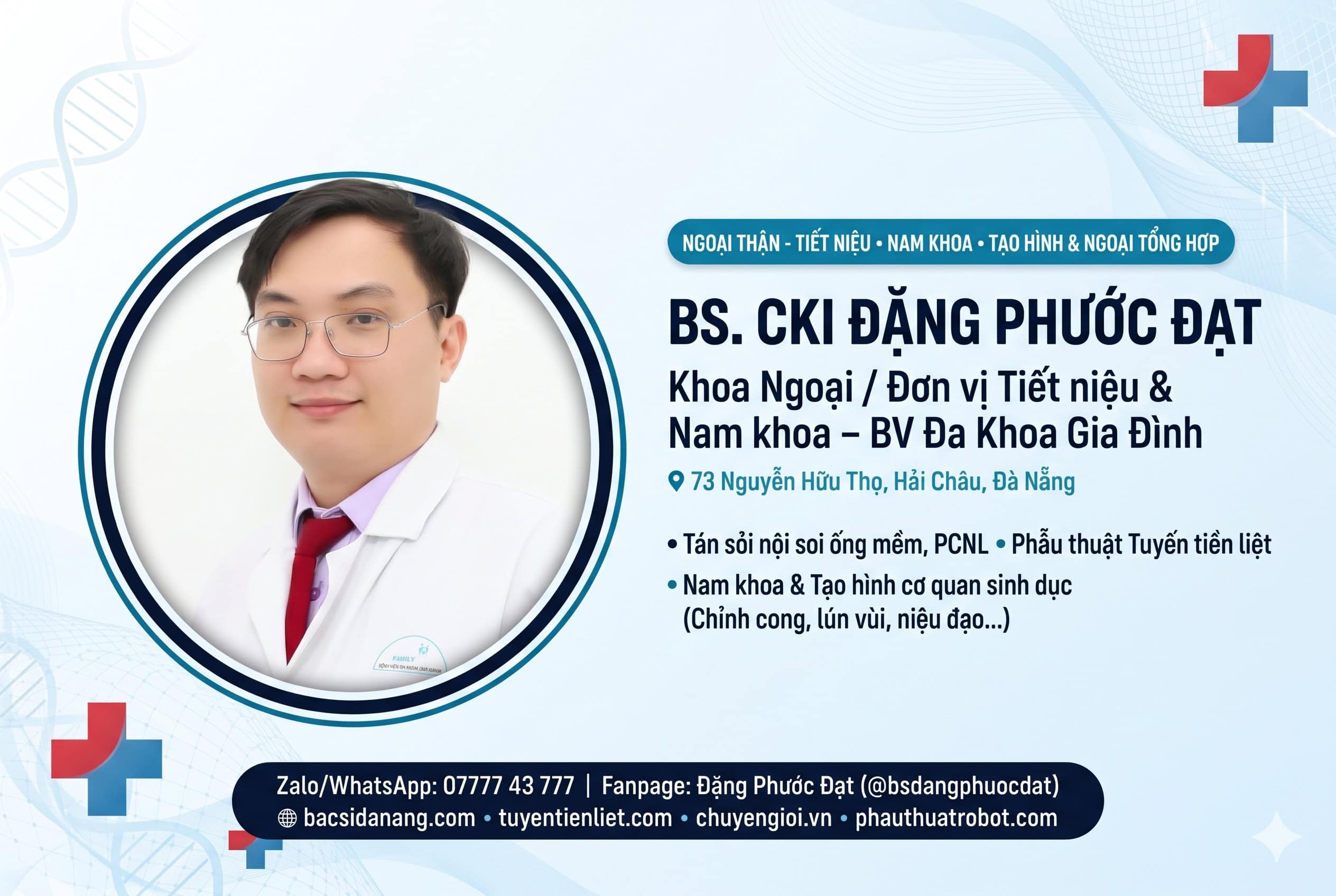
Author & Medical Reviewer: Dr. Dang Phuoc Dat, MD, Specialist Level I in Urology, Andrology & Microsurgery (Department of Surgery – Family Hospital Da Nang)
Executive Clinical Summary: Clinical varicocele (abnormal dilation and tortuosity of the pampiniform venous plexus within the spermatic cord) is the single most common correctable cause of male infertility worldwide—present in 35%–40% of men with primary infertility and up to 75%–80% of men with secondary infertility—as well as a frequent cause of dull, aching scrotal pain after standing or exercise. According to the European Association of Urology (EAU) and American Urological Association (AUA) guidelines, Microsurgical Subinguinal Varicocelectomy (Marmar–Goldstein technique) under high-power optical magnification is the undisputed gold standard, achieving a 60%–80% improvement in semen parameters, significant increases in serum testosterone, < 1% recurrence rate, and near-zero risk of postoperative hydrocele or testicular artery injury for patients in Da Nang, Vietnam.
1. How Does a Clinical Varicocele Impair Sperm DNA Integrity and Testosterone Production?
In healthy scrotal anatomy, the pampiniform venous plexus acts as a counter-current heat exchanger that cools arterial blood entering the testis to 2°C–3°C below core body temperature. When venous valves in the internal spermatic vein become incompetent (occurring on the left side in 85%–90% of cases due to the perpendicular insertion of the left gonadal vein into the high-pressure left renal vein), warm venous blood refluxes into the scrotum [1, 2]:
- Scrotal Hyperthermia & Oxidative Stress (ROS): Elevated intratesticular temperature triggers excessive reactive oxygen species (ROS), causing severe Sperm DNA Fragmentation (SDF) and oligoasthenoteratozoospermia (OAT syndrome: low count, poor motility, abnormal morphology) [3].
- Leydig Cell Dysfunction & Low Testosterone: Chronic venous stasis impairs Leydig cell steroidogenesis; published studies confirm that microsurgical varicocele repair increases serum total testosterone by 3.0–5.0 nmol/L (~85–140 ng/dL) [4].
CLINICAL GRADING & SURGICAL INDICATIONS FOR VARICOCELECTOMY (DUBIN–AMELAR & EAU GUIDELINES)
| Clinical Grade & Color Doppler Ultrasound | Physical Exam & Hemodynamic Findings | Evidence-Based Management by Dr. Dang Phuoc Dat |
|---|---|---|
| Subclinical Varicocele (Veins < 2.5–3.0 mm) | Not palpable on physical examination even with Valsalva maneuver; detected only on ultrasound. | Conservative management; surgery is not indicated for isolated subclinical varicocele [2]. |
| Clinical Grade I – II Varicocele (Veins ≥ 3.0 mm with reflux > 2 sec) | Palpable during Valsalva (Grade I) or palpable at rest without Valsalva (Grade II), accompanied by abnormal semen analysis or chronic scrotal pain. | Microsurgical Subinguinal Varicocelectomy when associated with abnormal semen parameters, elevated DNA fragmentation, or persistent orchialgia [5, 6]. |
| Clinical Grade III Varicocele (“Bag of Worms”) | Visibly dilated venous plexus through scrotal skin, often with ipsilateral testicular hypotrophy (> 20% volume loss). | Prompt Microsurgical Subinguinal Repair to arrest progressive testicular atrophy and restore spermatogenesis & Leydig function [1]. |
2. 4-Column Comparison: Microsurgical Subinguinal (Marmar) vs. Laparoscopic vs. Open Inguinal Repair
| Surgical Parameter | Microsurgical Subinguinal (Marmar Gold Standard) | Laparoscopic Varicocelectomy | Conventional Open Inguinal (Ivanissevich) |
|---|---|---|---|
| Anatomical Incision & Fascial Integrity | 2 cm mini-incision below external inguinal ring; external oblique aponeurosis is NOT cut (minimal pain) [5]. | 3 abdominal trocar incisions under general anesthesia. | 4–5 cm groin incision splitting the external oblique muscle fascia. |
| Spontaneous Pregnancy Rate | 42% – 47% within 12 months (+143% increase in motile sperm count) [1]. | 30% – 33%. | 28% – 35%. |
| Varicocele Recurrence Rate | < 1.0% (0.8% – 1.2%) due to ligation of both internal and external spermatic veins [1]. | 4% – 11% (cannot ligate external spermatic veins). | 9% – 15%. |
| Postoperative Hydrocele Risk | 0% – 0.4% (10x–20x optical magnification preserves all lymphatic vessels) [1]. | 5% – 12% hydrocele formation. | 7% – 10% hydrocele formation. |
3. Why Optical Magnification Is Critical During Subinguinal Varicocelectomy in Da Nang
At the subinguinal level just below the external inguinal ring, the internal spermatic artery measures only 0.5 to 1.0 mm in diameter and is tightly surrounded by a network of small adherent veins (venae comitantes) and delicate transparent lymphatics [5]. During surgery at Family Hospital Da Nang, Dr. Dang Phuoc Dat, MD, Specialist Level I utilizes high-magnification microsurgical dissection and topical papaverine microvascular vasodilation to:
- Clearly identify and preserve the pulsating testicular artery, cremasteric artery, and deferential artery.
- Isolate and spare 4 to 6 lymphatic channels, completely preventing postoperative scrotal hydrocele [1].
- Individually ligate and divide all dilated internal spermatic veins and external spermatic perforators while keeping the vas deferens and its vessels untouched.
⚠️ CLINICAL RED FLAGS IN VARICOCELE EVALUATION
Prompt abdominal/retroperitoneal CT or ultrasound imaging is mandatory if a patient presents with: (1) An isolated right-sided varicocele; (2) A sudden-onset varicocele in a man over 40 years of age; or (3) A large varicocele that fails to collapse when lying flat (supine position)—as these signs require ruling out retroperitoneal mass compression or renal cell carcinoma thrombus in the renal vein.
4. Frequently Asked Questions (FAQ) About Microsurgical Varicocelectomy
Q1: How soon after microsurgical varicocelectomy will my sperm count improve?
Answer: Because the human spermatogenic cycle takes approximately 72 to 74 days, follow-up semen analyses are performed at 3 months and 6 months postoperatively. Significant improvements in sperm concentration (+12 to +18 million/mL), progressive motility, and DNA fragmentation index are typically observed by month 3 to 6 [2, 5].
Q2: What is the recovery timeline for work, gym workouts, and sexual activity?
Answer: Because the subinguinal approach does not cut any abdominal muscle or fascia, patients are discharged within 24 hours, return to desk work in 2–3 days, resume sexual activity after 10–14 days, and return to heavy weightlifting or running after 3–4 weeks.
Medical References (Vancouver Style)
- Chan P. Management options of varicoceles. Indian J Urol. 2011;27(1):65-73. (NCBI PMC)
- Choi WS, Kim SW. Current Issues in Varicocele Management: a Review. World J Mens Health. 2013;31(1):12-20. (NCBI PMC)
- Pagani RL, Ohlander SJ, Niederberger CS. The role of microsurgical varicocelectomy in treating male infertility. Transl Androl Urol. 2017;6(Suppl 4):S322-S326. (NCBI PMC)
- Microsurgical Subinguinal Varicocele Repair of Grade II-III Lesions Improves Serum Testosterone Levels. Urol Int / PMC. 2017. (NCBI PMC)
- Lee HS, Park SH, et al. Microsurgical Intermediate Subinguinal Varicocelectomy. Int Neurourol J / PMC. 2014;18(2):97-102. (NCBI PMC)
- Anthropometric Variables as Predictors of Semen Parameters Following Subinguinal Varicocelectomy. Urol J / PMC. 2020. (NCBI PMC)
Select any symptoms you are currently experiencing to see your recommended clinical triage level before messaging Dr. Dang Phuoc Dat:

ĐẦU TƯ NGHIÊM TÚC