

 & staghorn stones via a 5mm keyhole. Consult Dr. Dang Phuoc Dat: +84919480180.)
Author & Medical Reviewer: Dr. Dang Phuoc Dat (Specialist Level I) – Urology, Andrology & Reconstructive Surgery, Family Hospital Da Nang | Updated: October 22, 2026
For large renal pelvic stones exceeding $20\text{ mm}$ or complex staghorn calculi branching across multiple calyces, patients once faced invasive open flank surgery with a 15–20 cm muscle-splitting incision. Today, mini PCNL kidney stone surgery Da Nang (Mini-Percutaneous Nephrolithotomy) serves as the international gold standard, achieving complete stone clearance through a single $5\text{ mm}$ keyhole puncture.
Quick Clinical Summary: Mini-PCNL creates a miniature $5\text{ mm}$ (16Fr–18Fr) percutaneous tunnel from the flank directly into the target renal calyx under real-time ultrasound guidance, utilizing high-power Holmium laser energy and hydrodynamic suction to clear large stones in a single session. Following EAU and AUA guidelines, Dr. Dang Phuoc Dat performs Ultrasound-Guided Mini-PCNL in Da Nang as first-line therapy for all renal stones $> 20\text{ mm}$, staghorn calculi, and complex lower-pole stones $> 15\text{ mm}$.

1. What Is Mini-P>1. What Is Mini-PCNL and How Does It Improve Upon Standard PCNL?
lock-paragraph">Conventional Standard PCNL requires a wide 24Fr–30Fr ($8–10\text{ mm}$) access sheath, which carries a higher risk of parenchymal bleeding. The evolution to Mini-PCNL downsizes the metallic working sheath to 16Fr–18Fr (approximately $5\text{ mm}$), delivering major clinical advantages:- Maximal Nephron & Vascular Preservation: Cross-sectional parenchymal trauma is reduced by more than 50%, entering precisely along the longitudinal axis of the renal papilla through Brodel’s Bloodless Line.
- Hydrodynamic ‘Vacuum Cleaner Effect’: Based on the Bernoulli principle, continuous irrigation between the slender nephroscope and the 16Fr–18Fr sheath generates a low-pressure vortex that automatically flushes crushed stone fragments out through the sheath without repeated grasping forceps passes.
Clinical Comparison: Ultr>Clinical Comparison: Ultrasound-Guided Mini-PCNL vs. Open Kidney Stone Surgery
k-table">| Clinical Criterion | Mini-PCNL Keyhole Surgery (16Fr–18Fr) | Traditional Open Nephrolithotomy |
|---|---|---|
| Incision Size | Single $5\text{ mm}$ skin puncture in the flank (pen-tip size) | 15 – 20 cm flank incision cutting through multiple abdominal/back muscle layers |
| Renal Parenchymal Impact | Minimal (< 1% of renal cortical surface), preserving eGFR | Requires extended pyelotomy or nephrotomy, risking cortical scarring |
| Stone-Free Rate (SFR) | 90% – 96% via direct nephroscopic inspection of individual calyces | Prone to residual fragments in hidden anterior/posterior calyces |
| Postoperative Pain & Stay | Mild discomfort, ambulation within 24 hours, discharge in 3 – 4 days | Significant wound pain, 7 – 10 day hospitalization, risk of incisional hernia |
2. Clinical Indications for Mini-PCNL in Da Nang
2. Clinical Indications for Mini-PCNL in Da NangDr. Dang Phuoc Dat stratifies stone complexity using the Guy’s Stone Score on preoperative CT Urography and recommends Mini-PCNL for:
- Large Renal Calculi $> 20\text{ mm}$ or partial/complete Staghorn Calculi occupying the renal pelvis and calyces.
- Lower-Pole Renal Stones $15–20\text{ mm}$ with unfavorable anatomy (narrow infundibulum $< 5\text{ mm}$ or acute infundibulopelvic angle $< 30^\circ$) where ESWL or flexible ureteroscopy cannot clear fragments.
- Large Impacted Proximal Ureteral Stones ($> 15\text{ mm}$) associated with Grade II–III hydronephrosis.
- Recurrent Stones in Previously Operated Kidneys or calculi refractory to ESWL/RIRS.
3. Radiation-Free Ultrasound-Guided Puncture Technique by Dr. Dan>3. Radiation-Free Ultrasound-Guided Puncture Technique by Dr. Dang Phuoc Dat
Phuoc Dat’s Mini-PCNL technique in Da Nang is 100% Real-Time Ultrasound-Guided Renal Access, eliminating ionizing X-ray fluoroscopy exposure for both patient and surgical team:- Step 1 (Retrograde Ureteral Catheterization): Under general anesthesia, a ureteral catheter is placed cystoscopically to instil sterile saline, gently distending the collecting system (artificial hydronephrosis) for pinpoint targeting.
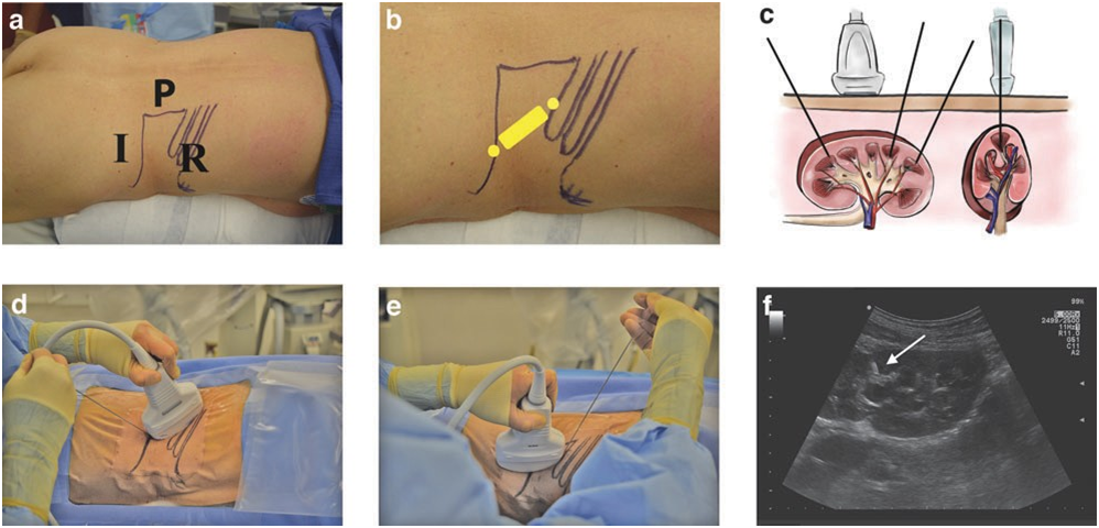
- Step 2 (Doppler Ultrasound Papillary Puncture & 5 mm Dilation): Color Doppler ultrasound visualizes intrarenal segmental arteries and adjacent organs (pleura, colon, spleen/liver), guiding the puncture needle directly through the target calyx fornix before dilating to 16Fr–18Fr.
- Step 3 (High-Power Holmium Laser Lithotripsy & Vacuum Evacuation): The miniature nephroscope fragments the stone burden with a 550 $\mu$m Holmium laser fiber while continuous hydrodynamic flow evacuates every fragment.
- Step 4 (Double-J Stenting & Tubeless Option): An internal Double-J Ureteral Stent is deployed anterogradely, with selected uncomplicated cases completed as Tubeless Mini-PCNL (no external nephrostomy bag).
Clinical Red Flags: When to Seek Immediate Urological Care
<>Clinical Red Flags: When to Seek Immediate Urological Carer:#fff5f5;border-left-color:#d32f2f;border-left-width:4px;padding-top:16px;padding-right:20px;padding-bottom:16px;padding-left:20px">URGENT MEDICAL RED FLAGS:
- During the first 7–14 days after discharge following Mini-PCNL, patients must avoid heavy lifting, strenuous exercise, or vigorous running to allow complete healing of the parenchymal tract.
- Seek urgent urological evaluation if you develop bright red hematuria with blood clots, flank swelling, or high fever with chills.